Contrast ventriculography is a specialized imaging technique that uses radiopaque or paramagnetic contrast agents injected directly into the heart's ventricles or the brain's ventricular system to evaluate structure, function, and fluid dynamics. This procedure provides critical diagnostic information that standard imaging often cannot achieve with the same level of detail.
There are two primary contexts for contrast ventriculography: cardiac ventriculography (most commonly left ventricular) and cerebral (MR) ventriculography. Both rely on contrast media to opacify chambers or spaces for X-ray, fluoroscopic, or magnetic resonance imaging.
Cardiac Ventriculography
Cardiac ventriculography, often performed during cardiac catheterization, involves injecting an iodinated contrast agent into the left or right ventricle. In left ventriculography, a catheter is advanced across the aortic valve into the left ventricle, and contrast is injected rapidly while cineangiography records the heart's contraction. This allows clinicians to assess:
· Ejection fraction (EF): The percentage of blood pumped out of the left ventricle with each heartbeat (normal >50-55%).
· Wall motion abnormalities: Such as hypokinesis, akinesis, or dyskinesis, which indicate prior myocardial infarction or ischemia.
· Ventricular volumes: End-diastolic and end-systolic volumes.
· Structural issues: Including mitral regurgitation, ventricular septal defects, or aneurysms.
· Right ventriculography evaluates the right ventricle, pulmonary valve, and tricuspid valve function. The procedure is typically combined with coronary angiography and provides real-time functional data during invasive cardiac procedures.
Advantages include high spatial and temporal resolution for dynamic assessment of ventricular contraction. However, modern non-invasive alternatives like echocardiography, cardiac MRI, and nuclear ventriculography (MUGA scan) have reduced routine use. Studies show left ventriculography is frequently performed even when recent non-invasive EF measurements exist, raising concerns about overuse due to added contrast load, radiation, and procedural risks.
Cerebral MR Ventriculography
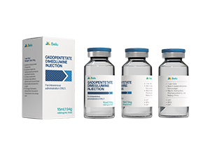
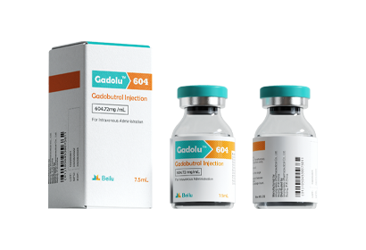
In neuroimaging, MR ventriculography uses gadolinium-based contrast agents injected directly into the cerebral ventricles via an existing ventricular drain or puncture to study cerebrospinal fluid (CSF) flow dynamics. This is particularly valuable in patients with hydrocephalus, suspected obstructions, arachnoiditis, or intraventricular lesions.
By injecting a small volume of diluted gadolinium contrast, radiologists can track flow through the lateral ventricles, foramina of Monro, third and fourth ventricles, and outlets into the subarachnoid space. It helps identify blockages, cysts, or leptomeningeal disease that may not be fully characterized on standard MRI or CT.
Procedure and Patient Preparation
For cardiac ventriculography, patients undergo preparation similar to coronary angiography: fasting, renal function assessment, and allergy history review. The procedure occurs in a catheterization lab under local anesthesia and mild sedation. Contrast injection is power-controlled to avoid complications.
MR ventriculography requires neurosurgical access to the ventricles and is performed in an MRI suite. Patients must be stable enough for transport and imaging.
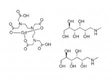
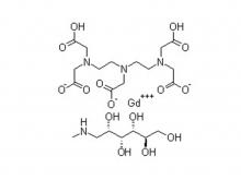
Iodinated contrast agents, non-ionic, low- or iso-osmolar, are standard for cardiac applications, while gadolinium-based agents are used for MR ventriculography due to their paramagnetic properties that enhance T1-weighted images.
Risks and Safety Considerations
· Cardiac: Additional contrast exposure (increasing risk of nephropathy, especially in diabetic or renally impaired patients), arrhythmias, stroke from catheter manipulation or emboli, and rare allergic reactions.
· Cerebral MR: Potential for increased intracranial pressure, infection, or gadolinium-related issues, though diluted intrathecal use is generally well-tolerated in controlled settings.
· General: Radiation exposure, bleeding at access sites, and contrast reactions.
· Pre-hydration, minimal effective dosing, and patient selection minimize these risks. Non-ionic low-osmolar agents have substantially improved safety over older high-osmolar formulations.
Beilu Pharma's Role in Contrast Ventriculography
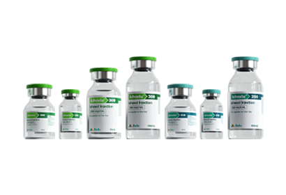
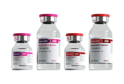
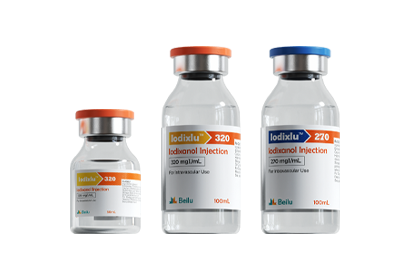
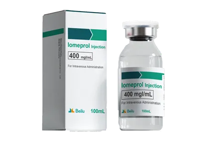
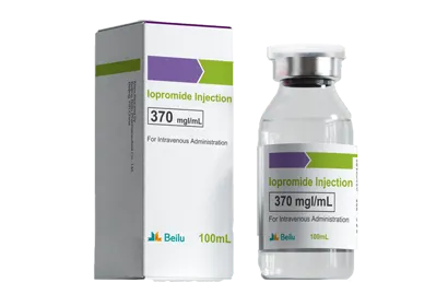
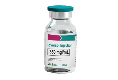
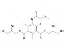
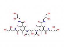
Beilu Pharma provides high-quality contrast media suitable for ventriculography procedures. As a pioneer in China's contrast media products industry, Beilu offers a comprehensive range of iodinated agents, such as Iopamidol, Iohexol, and Iodixanol, ideal for ventriculography. Their products are manufactured under stringent EU GMP standards, ensuring reliability for both diagnostic and interventional use.
Beilu Pharma's formulations support clear visualization of ventricular function and CSF dynamics, contributing to accurate diagnosis in complex cardiac and neurological cases. The company's commitment to quality has made its contrast agents trusted options in clinical settings worldwide.
Clinical Applications and Outcomes
Cardiac ventriculography remains valuable for detailed functional assessment before interventions like valve replacement or in cases where non-invasive imaging is inconclusive. Cerebral MR ventriculography excels in preoperative planning for hydrocephalus shunting or evaluating CSF flow obstructions.
Normal results show vigorous, symmetric contraction (cardiac) or unobstructed CSF flow (cerebral). Abnormal findings guide therapy, from revascularization in ischemic cardiomyopathy to surgical decompression in obstructive hydrocephalus.
Future Perspectives
Advances in imaging technology, such as 3D rotational angiography and AI-enhanced analysis, continue to refine ventriculography. While non-invasive methods dominate routine evaluation, contrast-enhanced ventriculography retains a key role in complex or intraoperative settings.
In summary, contrast ventriculography delivers irreplaceable insights into ventricular performance and CSF pathways. When performed with modern, low-risk agents, it supports precise diagnosis and improved patient management across cardiology and neuroradiology. Clinicians must balance its benefits against risks, reserving it for situations where it will meaningfully impact care.

 EN
EN
 jp
jp  fr
fr  de
de  es
es  ru
ru  pt
pt  ar
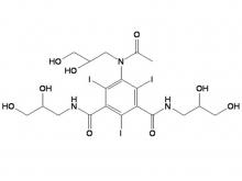
ar 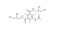 Iohexol Intermediate 5-Amino-N,N'-bis(2,3-dihydroxypropyl)-2,4,6-triiodo-1,3-benzenedicarboxamide
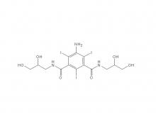 Iohexol/Ioversol Intermediate 5-Amino-N, N'-bis(2,3-dihydroxypropyl)-2,4,6-triiodo-1,3-benzenedicarboxamide
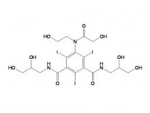
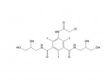 Ioversol Intermediate (order based) N, N'-Bis(2,3-dihydroxypropyl)-5-(glycoloylamino)-2,4,6-triiodoisophthalamide
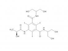
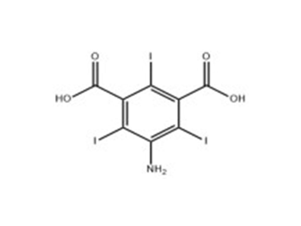
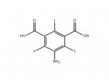 Iopamidol Intermediate (order based) 5-Amino-2,4,6-triiodoisophthalic acid
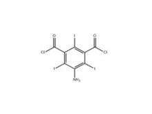 Iopamidol Intermediate (order based) 5-Amino-2,4,6- triiodisophthaloyl acid dichloride
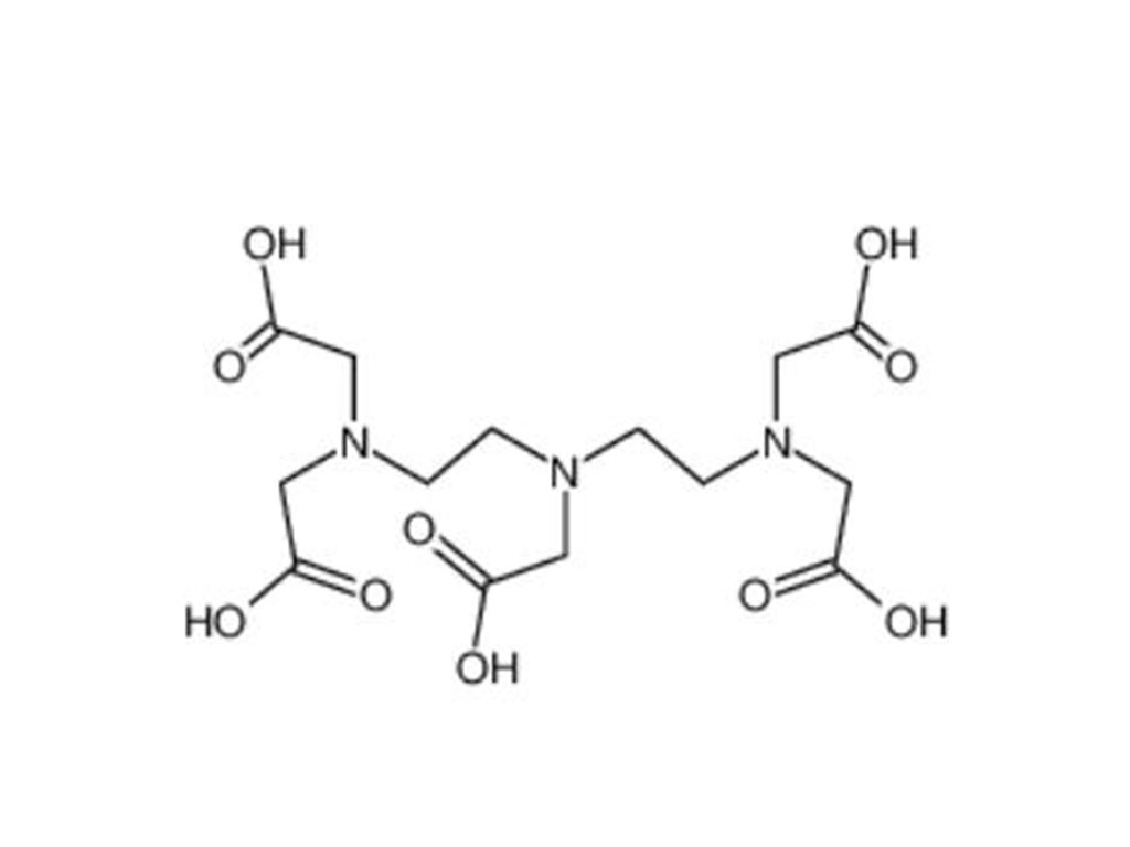
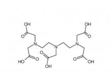 Diethylenetriaminepentaacetic acid (DTPA)




 Call us on:
  Email Us:
  No.3 Shuiyuan West Road, Miyun District, Beijing, China